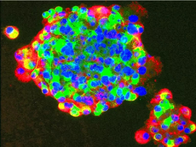
Very recently some Scientists have produced images of the novel coronavirus infecting lab-grown respiratory tract cells. They discovered new concept of cell and findings that illustrate the number of virus particles that are produced and released per cell inside the lungs.
The health researchers including Camille Ehre from the University of North Carolina (UNC) Children’s Research Institute. Their team of the scientist captured these images to illustrate how intense SARS-CoV-2 infection of the airways can be in very graphic and easily understood images.
Image of the damaged cell published in the New England Journal of Medicine, they were re-colorized and show infected hairy conciliated cells with strands of mucus attached to cilia tips.
After seeing the image scientists explained that the cilia are hair-like structures on the surface of airway epithelial cells that transport mucus and trapped viruses from the lungs.
As per Scientist the large viral burden is a source for spread of infection to multiple organs of an infected individual, and also its likely mediates the high frequency of Covid-19 transmission to others.

![Buddha Purnima 2025 [TKB INDIA]](https://topknowledgebox.com/iphaphoo/2025/05/12052025-150x150.jpg)
![YouTube is about to turn 20, the company announced many big features [TKB Tech]](https://topknowledgebox.com/iphaphoo/2025/04/28042025-150x150.jpg)
![Basant Panchami 2025: Know the correct date and auspicious time [TKB INDIA]](https://topknowledgebox.com/iphaphoo/2025/01/31012025-150x150.jpg)

![Amazing feature of WhatsApp, you will be able to reply without listening to the voice message[TKB Tech]](https://topknowledgebox.com/iphaphoo/2024/11/24112024-150x150.jpg)


More Stories
Essential Oil For Skin[TKB Health]
Chia seeds will give double benefits [TKB Health]
Sunglasses Choosing Tips: Keep note before buying sunglasses, otherwise your eyes may get damaged! [TKB Health]